
The impact of anthracosis on nuclei segmentation in lung tissue
Aim
Anthracosis are dust particles which get accumulated within the stroma and macrophages in lung tissue. These particles can be found in all lungs but are particularly frequent in smokers. On histological slides, anthracosis is visible as black pigments both in the classical H&E and immunohistochemical staining. Thus, anthracotic pigments have a high optical density and get typically segmented when using threshold- and deep-learning-based methods to detect nuclei. As it is an organ-specific artefact most publicly available datasets do not include examples of anthracosis. Furthermore, there is a lack of public immunohistochemical datasets challenging the use of available segmentation methods in the routine diagnostic workflow. However, it is especially in these slides that anthracosis will affect the downstream analyses as these pigments get typically detected as antibody-positive nuclei.
In this project, we aim to efficiently and accurately segment anthracotic pigments in CD8-stained slides of non-small cell lung cancer cases. The segmentation will be used to quantify anthracosis in the slide and exclude false positive nuclei from further downstream analyses.
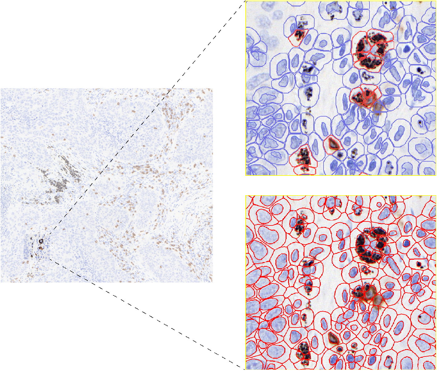
Figure 1. Classic approaches such as watershed segmentation and convolutional neural network-based models fail to neglect anthracotic pigments during nuclei segmentation.

Figure 2. Segmentation of anthracotic pigments used for quantification and negative masking.
Members

Philipp Zens

Sabina Berezowska